
Welcome
The Department of Radiology at NuHealth offers an approved four-year program in Diagnostic Radiology, leading to board certification. The program received a five year accreditation in 2008, with no citations. The 26-room department performs more than 150,000 examinations a year in all Diagnostic Radiology subspecialty areas. Our department is under the supervision of a dedicated, full-time staff. Our hospital is in the process of a major modernization effort, and our department has benefited with new state-of-the-art equipment for most areas.
A newly renovated departmental library/lounge, including a teaching file on DVD and other audiovisual aids, permits ready access to current and standard educational materials. A large conference room with audiovisual equipment is located within the department. Four Residency positions are offered each year.
Follow this link to meet our Residents and take a tour of our Department www.numcradiology.com
The resident board’s pass rate was 100% in the June 2014 ABR examinations. The residents are productive with scholarly activities, exhibits, oral presentations and publications in national meetings such as Radiological Society of North America, American Roentgen Ray Society and American Society of Neuroradiology. The activities are in the fields of clinical radiology and quality improvement. Here is the latest list of scholarly activities.
We maintain a program in which Residents learn in a comfortable, upbeat, and progressive environment. We provide top-quality patient care while training outstanding clinical and academic Radiologists. Our faculty members are dedicated to promoting a rich environment for learning, teaching, and practicing clinical radiology. To promote and encourage Resident input in department matters, Residents participate in departmental meetings and have a seat in many hospital committees. Residents have been integral in protocol development, lecture and curriculum changes as well as IT initiatives.
Our Residency curriculum is structured, and includes several rotations at our affiliated hospitals in the North Shore-Long Island Jewish Health System. These outside rotations supplement case volume in areas such as cardiac radiology, pediatric radiology and PET imaging.
The department conducts a comprehensive teaching program to prepare Residents for their board certification. Lectures on physics are part of the Residency program. The Physicists are also available for consultation on issues related to radiation safety. At the end of their training, our Residents become independent, confident and highly competent Radiologists, whether they choose private practice or an academic career.
We welcome your interest in our Residency program, and we look forward to seeing you soon.
Steven Lev, MD
Chairman, Department of Radiology
Salman Shah, MD
Program Director
Program Description
Radiology encompasses a variety of diagnostic and image-guided therapeutic techniques, including all aspects of radiological diagnosis, nuclear radiology, diagnostic ultrasound, magnetic resonance, computed tomography, interventional procedures, and the use of other forms of radiant energy. Our Residency program offers a quality graduate medical educational experience of adequate scope and depth in all of these associated diagnostic disciplines. We offer an environment that encourages the interchange of knowledge and experience among Residents and faculty in the program, and with Residents and faculty in other major clinical specialties throughout the hospital.
CURRICULUM OVERVIEW
DIAGNOSTIC RADIOLOGY
The Division of Diagnostic Radiology offers a broad experience in the field. The large number of patients seen at the institution, particularly in the Emergency Department, provides extensive exposure to trauma radiology. The Radiology staff includes specialists in GI, GU, Chest, Bone, Ultrasound, Body CT/MRI, Nuclear Medicine, Mammography, Vascular and Interventional Radiology, and Neuroradiology.
In all areas, Residents are given increasing responsibility for the performance and interpretation of radiographic examinations under the direct supervision of an Attending Radiologist. There are multiple intra-departmental and interdepartmental educational conferences to promote learning.
NEURORADIOLOGY
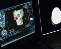
The Division of Neuroradiology is responsible for all neuroradiological procedures, including computed tomography, magnetic resonance imaging, cerebral angiography, and myelography. There is one 16-slice and one 240-slice MDCTs, a 1.5 T magnetic resonance imaging unit, and a digital angiography suite for imaging of the brain and spine. The Residents who rotate through the Division of Neuroradiology actively participate in the performance and interpretation of all neuroradiological procedures under the guidance of a highly qualified Neuroradiologist. The assigned Residents also help prepare and actively participate in all intradepartmental and interdepartmental conferences pertaining to Neuroradiology.
ULTRASOUND, BODY COMPUTED TOMOGRAPHY AND MAGNETIC RESONANCE IMAGING
The Divisions of Ultrasound, Body Computed Tomography, and Magnetic Resonance Imaging are part of the Department of Radiology. There is a close interaction between these diagnostic imaging modalities.
There is a large obstetrical referral service which refers patients for ultrasound evaluation of high-risk pregnancies. Ultrasonography is also used in the diagnosis of pelvic, abdominal, and vascular abnormalities, as well as evaluation of other organ systems. All ultrasound examinations are performed with state-of-the-art equipment, including color, pulsed and power Doppler. Scanning techniques and interpretation are both stressed.
Two state-of-the-art CT scanners and a newly upgraded magnetic resonance imaging unit provide the entire gamut of cross-sectional imaging examinations. There is a state-of-the-art 320-slice CT scanner within the newly expanded Emergency Department to increase the services offered, including cardiac CT, abdominal, pelvic, chest, and musculoskeletal MRI examinations which are performed. Experience in body CT is extensive, including ample exposure to trauma studies. Residents also participate in the planning and performance of CT and ultrasound-guided biopsy and drainage procedures. During their rotations in body-imaging divisions, Residents gain considerable experience in planning, performing, and reviewing cases, with one-on-one interaction with Attending Radiologists. Daily teaching is given to Residents by the Attending staff. Additional learning and discussion is provided through interesting case and interdepartmental conferences.
BREAST IMAGING
The Division of Breast Imaging offers extensive experience in the diagnosis of breast diseases. The Breast Imaging Center has three dedicated mammography units, and a dedicated breast ultrasound unit. There is an increasing volume of breast MRI examinations.
Residents participate in the planning and performance of interventional procedures, including needle localizations and biopsies. Biopsies are performed under MRI, ultrasound and stereotactic localization. The Resident works closely with the Attending staff, assuming increasing responsibility for patient evaluation and treatment.
NUCLEAR MEDICINE
Radionuclide in vivo imaging and laboratory examinations are performed in a separate suite containing state-of-the-art equipment. Two gamma cameras, as well as state-of-the-art SPECT/CT, are used for general as well as nuclear cardiology studies. A dual-energy absorptiometry (DEXA) unit is used for osteoporosis detection.
The Residents’ primary responsibility is not only to interpret all in vivo studies, but to evaluate every request for a nuclide procedure. Patient charts, as well as all prior X-Ray, ultrasound and CT studies, are immediately available to aid in formulating an impression and guiding the clinician to the next diagnostic step. Each study is reviewed before the patient leaves the imaging suite. Scans are reviewed by the Attending staff for consultation and confirmation of the trainee’s impression.
Regularly scheduled conferences are held within the Department of Radiology, as well as in conjunction with other services. Clinical conferences are held in the division itself.
VASCULAR AND INTERVENTIONAL RADIOLOGY
The Division of Vascular and Interventional Radiology offers the full range of outpatient and inpatient special procedures. These include arteriography and venography; peripherally inserted central catheters (PICCS); placement of ports in the upper arm; venous sampling procedures; lytic therapy; balloon angioplasty and stent placement for arterial, venous, or graft stenosis; embolizations; hepatobiliary procedures such as transhepatic cholangiograms; biliary drainage and dilatation; percutaneous nephrostomies; abscess drainage; and biopsies of various organs. Equipment includes two angiography suites, one of which has digital capability. Residents assume gradually increasing responsibility for these procedures, under the guidance of their Attending, as their experience increases. Weekly teaching conferences are held in the department.
We are proud to offer Early Specialization in Interventional Radiology (ESIR) pathway to interested residents. Our institution is accredited for one to two residents per year. After completing IR rotations in first year of residency, applicants apply for ESIR in their second year of residency.
RADIATION ONCOLOGY
The Division of Radiation Oncology at NuHealth is a modern cancer treatment center. Equipment includes a state-of-the-art high-dual-energy linear accelerator with electrons and a cobalt unit. A new 3-D planning system and IMRT-capability have just been installed. Brachytherapy procedures with radioactive materials are performed in conjunction with CT scans.
With over 10,000 patients per year, the division participates in a full-scale, integrated, multi-disciplinary cancer treatment program with other services.
DIVISION OF PHYSICS
The Division of Physics of the Department of Radiology provides direct support to the clinical divisions and oversees the radioactive materials and X-ray radiation safety programs for the entire medical center.
The division maintains an exemplary quality-assurance program. The division utilizes two computers for clinical purposes, one dedicated to the Division of Nuclear Medicine for the purpose of data acquisition and analysis, and the other to the Division of Radiation Oncology for determination of patient dose distributions and other numerical processing. An extensive range of software is utilized, and Resident physicians are encouraged to interact with both systems.
Faculty
Steven Lev, MD, Chairman, Department of Radiology
Medical School: Mount Sinai School of Medicine
Residency: Diagnostic Radiology, Beth Israel Medical Center
Fellowship: Neuroradiology, New England Medical Center, Tufts University School of Medicine
Salman Shah, MD, Chief, Vascular and Interventional Radiology,
Medical School: State University of New York College of Medicine
Residency: Diagnostic Radiology, Mount Sinai School of Medicine, Chief Resident
Fellowship: Vascular and Interventional Radiology, Mount Sinai School of Medicine, Chief Administrative Fellow
Susan Gottlieb, MD, Attending
Medical School: The Mount Sinai School of Medicine
Residency: Radiology, Albert Einstein College of Medicine Montefiore Medical Center
Fellowship: Mammography, New York University Medical Center US, CT & MRI, Thomas Jefferson University Hospital
John Krumenacker, MD, Chief, Pulmonary Radiology
Medical School: SUNY Stony Brook
Residency: Diagnostic Radiology, New England Medical Center, Tufts University School of Medicine
Fellowship: Body Imaging, Memorial Sloan-Kettering Cancer Center
Shari Lobel, MD, Attending
Medical School: SUNY Downstate Medical School
Residency: Diagnostic Radiology, Long Island College Hospital
Fellowship: Abdominal & Obstetrical US, CT & MR, New York Hospital, Cornell University Medical Center
Joel Rosen, MD, Chief, Nuclear Medicine
Medical School: Cornell University Medical College
Residency: Diagnostic Radiology, SUNY Downstate Medical Center
Fellowship: Nuclear Medicine, Columbia-Presbyterian Medical Center; Imaging, Mount Sinai Medical Center
Ephram Weingarten, MD, Chief, Breast Imaging
Medical School: Mount Sinai School of Medicine
Residency: Diagnostic Radiology, Mount Sinai Medical Center
Fellowship: Vascular & Interventional Radiology, SUNY at Stony Brook
Mammography and General Sonography, Medical University of South Carolina
Dahua Zhou, MD, Chief, Ultrasound
Medical School: China Medical University
Residency: Diagnostic Radiology, Nassau University Medical Center; Radiation Oncology, China-Japan Friendship Hospital
Fellowship: Body Imaging, Memorial Sloan-Kettering Cancer Center
Useful Links
- American Board of Radiology (ABR)
- American College of Radiology (ACR)
- Radiological Society of North America (RSNA)
- Society of Breast Imagining
- Society of Computed Body Tomography & Magnetic Resonance
- Society of Interventional Radiology (SIR)
- Society of Nuclear Medicine
- Society of Radiologists in Ultrasound
Contact Us
Melissa Goldsmith
Program Coordinator
Telephone: 516 572-6785
Fax Number: 516-572-5941
Email: mgoldsmi@numc.edu
Salman Shah, MD
Program Director
Telephone: 516 572-6633
Email: sshah4@numc.edu
Other Important Information
What are the application deadlines?
The deadlines and general information can be obtained from the FRIEDA website, or by calling 516-572-6785.
When do you interview?
Interviews start in November, and continue through the end of January.
What is the format for the interview day?
You will have at least three formal interviews by the Program Director, the Chair, and one of the Chief Residents. Lunch will be provided, and you will attend a noon conference. You will have an opportunity to meet with many of the residents informally during your interview day. You will tour our department, and be able to observe our normal workday. The interview session typically starts at 9 AM and ends around 2 PM.
What is the format for the academic year?
Residents rotate at NUMC in the following areas within the department of Radiology: Body CT, Ultrasound, Body MRI, Chest, ICU, ER/Musculoskeletal, Pediatrics, GI/GU, Interventional Radiology, Neuroradiology and Nuclear Medicine. Residents go to LIJ for rotations in Pediatrics and Nuclear Medicine. Residents also go to North Shore Manhasset for rotations in Cardiac, Ultrasound and Interventional and Nuclear Medicine. The Breast Imaging rotation takes place in our newly renovated Breast Imaging Center. All residents go to AFIP for 4 weeks during the third year. Tuition for the course is paid by the department.
What is the call schedule format?
There is a night float system, which covers most of the overnight call. You do not begin to take call alone without either a senior resident or attending with you until your second year. Most times there is both a senior and junior resident in house.
Is there an opportunity for residents to perform research?
Our major focus is on learning clinical radiology, but there are ample opportunities for participation in research projects with many of the attendings. Most residents are involved with at least one project during their residency training, and some publish multiple papers.
Are residents able to go to conferences/academic meetings, and is there a book fund?
Residents are able to attend meetings at which their research is accepted for presentation. The department reimburses for the meeting and travel expenses. Residents are expected to publish research, which is presented at these meetings. Additionally, time and reimbursement for board-review courses for both the physics and oral boards is provided. There is no resident book fund. The department library is updated often.
Do you have PACS?
Yes. AMICAS PACS has been operational since June 2005, and we have a full online digital-image archive.
Is there elective time?
There is no elective time possible due to the varied nature of the schedule. Protected study time is permitted for board preparation.
How many residents are there?
There are 16 residents in the program, generally four per year.
Are there fellows?
There are no fellows in the department, so there are more than enough imaging studies and procedures for every resident.
Who can I contact for more information or to arrange a “second look” ?
You can contact the residency program coordinator at 516-572-6785.
ORAL PRESENTATIONS
First Place, NUMC’s Sixth Annual Research Day 2013:
RADIOLOGY
Improving the Workflow and Efficiency in a Breast Imaging Center: Decreasing Turn-Around Time for Diagnostic Mammography
Brandon Mirochnik, MD, Subah Gupta, MD, Steven Lev, MD
This was presented as an oral presentation at ARRS 2013 in Washington, DC, April 14-19, 2013
B. Goodman, 3rd Place Award for oral presentation at NUMC 7th Annual Graduate Medical Education Research Day, May 2014
RADIOLOGY & CARDIOLOGY
NASCI (North American Society for Cardiovascular Imaging)
Poster Presentation
9/27-10/1/2013 – 41st Annual NASCI Conference in Atlanta, Georgia
“Improving the Efficiency of the Stress Test”
Attendings:
Dr. Amgad Makaryus, Chairman of the Dept. of Cardiology
Dr. Steven Lev, Director of Neuroradiology
Residents:
Dr. Judy Atallah, DO, RY-4,
Dr. Subah Gupta, MD, RY-1
PUBLICATIONS
- Goodman B, Mirochnik B, Nagle J, Shah S. Improving the Efficiency of an Interventional Radiology Consultation Service: Abstract publication JVIR March 2014, Volume 25, Issue 3. Currently submitting the manuscript.
- S. Greenberg, N. Kanth, A. Kanth: A Woman with Cough: Gastrobronchial fistula as a delayed complication of bariatric surgery. Case report and literature review – The American Journal of Emergency Medicine online publication, 2013. http://www.ajemjournal.com/article/S0735-6757(13)00782-1/abstract
- Mirochnik BD, Weingarten EP. Digital Breast Tomosynthesis: A new Modality for Breast Cancer Screening. Contemporary Diagnostic Radiology. 36(14):1-5, July 1, 2013.
- Singh, K; Yakoub, D; Giangola, P; DeCicca, M; Patel, C; Marzouk, F; Giangola, G. Early follow-up and treatment recommendations for isolated calf deep venous thrombosis; 2012 Jan;55(1):136-140, Journal of Vascular Surgery.
- B. Mirochnik; P. Bhargava; M. Dighe; N. Kanth: Ultrasound Evaluation of Scrotal Pathology. Radiological clinics of North America 2012 Vol 50, no: 12;317-332.
- B. Mirochnik; P. Bhargava; M. Dighe; N. Kanth: Ultrasound Evaluation of Scrotal Pathology. In press, Radiological clinics of North America 2012.
PRESENTATIONS, EXHIBITS & ABSTRACTS
- Rubin A, Lev S. Back to the Basics with Building Blocks: A pattern based approach to teaching head CT, to be submitted to ASNR 2015.
- Cantos A, Yoon H, Goodman B. Reducing Fluoroscopy Time and Radiation Exposure During PICC Placement: To be submitted for 2015 annual meeting of the Society of Interventional Radiology (SIR).
- Holstad J, Macadam B, Zhou D. MDCT Findings of Soft Tissue Barotrauma: Striking, Subtle, and Mimics.
RSNA 2014 Educational Exhibit. - J. Holstad, American Journal of Neuroradiology: Fellows Journal Club Podcast, March 2014. Discussed journal article as 2nd-year resident.
- Caci D, Patel C, Khuong E, Shah S. The DR/IR Medical Student Clinical Rotation; Separate or Combined? Submitted: Society of Interventional Radiology 39th Annual Scientific Meeting; 2014 Mar 22-27.
- Shah S, Caci D, Fischman AM, Pessin E, Fung JW, Weintraub JL, Kim E, Nowakowski FS, Lookstein RA. Prospective Evaluation of the Safety and Efficacy of the Mynx Vascular Closure Device in Interventional Oncology Patients with Thrombocytopenia. Submitted: International Symposium on Endovascular Therapy 26th Annual Meeting; 2014 Jan 18-22.
- Rubin A, Byrns K, Dahua Z. Acute Pancreatic Injury and Associated Complications: The commonplace, the complex, and the curious, presented at ARRS 2014 Annual Meeting.
- Agrawal J, Gupta S, Gilani S, Lev S. Improving the PICC Line Workflow. In: Proceedings of the Society of Interventional Radiology, San Diego, CA, 2014.
- Goodman B, Atallah J, Lev S. The Delayed Complications of Brain Trauma: When the Worst is yet to Come. In: The American Roentgen Ray Society 2014 Annual Meeting, San Diego, CA 2014.
- Agrawal J, Lev S. Vision and Perception: A Teaching Module to Improve the Detection of Lesions on Head CT. Proceedings of the 52th Annual Meeting of the American Society of Neuroradiology, Montreal, QC, 2014.
- Holstad J, Rubin A, Lev S. Crime Scene Investigation, Neuroradiology Unit: Can you Solve the Case? In: Proceedings of the 52th Annual Meeting of the American Society of Neuroradiology, Montreal, QC, 2014.
- K. Byrns. Acute Pancreatic Injury and Associated Complications: The Commonplace, the Complex, and the Curious. ARRS American Roentgen Ray Society, 5/4/2014.
- Goodman B, Lev S. Delayed Complications in Brain Trauma: When the Worst is Yet to Come; Selected for Educational Exhibit at the American Roentgen Ray Society Annual Meeting, May 2014
- B. Goodman. Celiac Artery Dissection in Blunt Abdominal Trauma and Traumatic Abdominal Wall Hernia. 2014. Selected for Interesting Case Presentations at two different meetings of the Long Island Radiological Society.
- Caci, D; Patel, C; Khuong, E; Shah, S. Assessment of medical student awareness of the new Interventional Radiology and Diagnostic Radiology certification and the need for a separate core Interventional Radiology rotation. SIR annual meeting San Diego, CA (March 2014).
- Rubin, A; Byrns, K; Patel C; Zhou, D. Acute pancreatic injury and associated complications. ARRS annual meeting San Diego, CA (May 2014).
- K. Byrns. A View from Within – Virtual Flythrough Software as a Novel Method of Visualizing Head and Neck Pathology, RSNA – Radiological Society of North America, 12/1/2013.
- K. Byrns. False Alarm-A Primer on Differentiating Trauma from Mimics for the On-Call Radiologist.
RSNA – Radiological Society of North America, 12/1/2013. - Pan Q, Holstad J, Byrns K, Penna, Lev S. Would You Know it if it Hit You in the Face?
ASNR 2013 Educational Exhibit. - Jacob J, Byrns K, Artime C, Lev S. The Radiological Spectrum of Cranial Nerve Injuries in Head and Neck Trauma.
ASHNR – American Society of Head and Neck Radiology, ASHNR 47th Annual Meeting, 9/10/2013. - Byrns K, Rubin A, Lev S. Normal Variants and Mimics in Spinal Trauma Every Radiologist Should Know.
ASSR – American Society of Spine Radiology, Annual Symposium, 5/18/2013. - Byrns K, Lev S. A Walking Tour of the Temporal Bone.
ASNR – American Society of Neuroradiology, 51st Annual Meeting, 5/18/2013. - Pan Q, Holstad J, Byrns K, Penna K, Lev S. Exploring the Radiological Link Between Facial Fractures and Intracranial Hemorrhage. Presented at ASNR 51st Annual Meeting, 2013.
- B. MacAdam. Pandora’s Box – CT Findings following Cranial Surgery: Electronic Exhibit, ASNR 2013. Rehabilitation and Traumatic Brain Injury: Insights for the Neuroradiologist: Education Exhibit, ASNR.
- Caci D, Gilani S, Macadam B, Nelson K, Singhal R, Chowdry Y, Lev S. Rehabilitation and Traumatic Brain Injury: Insights for the Neuroradiologist [abstract]. In: American Society of Neuroradiology 50th Annual Meeting; 2013 Apr 21-25; New York, NY. 12-EdE-2180-ASNR. Abstract # 2180.
- Greenberg S, Byrns K, Atallah J, Krumenacker JH, Lev S. False Alarm – A Primer on Differentiating Trauma from Mimics for the On-Call Radiologist. In: Proceedings of the 99th Scientific Assembly and Annual Meeting of the Radiological Society of North America, Chicago, IL, 2013. 5
- Byrns K, Cunnane ME, Lev S. A View from Within – Virtual Flythrough Software as a Novel Method of Visualizing Head and Neck Pathology. In: Proceedings of the 99th Scientific Assembly and Annual Meeting of the Radiological Society of North America, Chicago, IL, 2013.
- Mirochnik B, Krumenacker J. Danger, Radiologist, Danger! Missed Lesions on Chest Radiograph. In: The American Roentgen Ray Society 2012 Annual Meeting, Vancouver, BC, 2012.
- Mirochnik B, Gottlieb S, Lobel S, Weingarten, E. Improving on a Good Thing: Breast Tomosynthesis. In: The American Roentgen Ray Society 2012 Annual Meeting, Vancouver, BC, 2012.
- Mirochnik B, Lev S. Becoming a Grandmaster: How Improving Your Chess Game Can Make You a Better Neuroradiologist. In: The American Society of Neuroradiology Annual Meeting, New York, NY, 2012.
- Nadendla P, Rubin A, Byrns K, Lev S. Ringing and Swishing, Humming and Buzzing: The Radiological Work-up of Tinnitus, presented at the ASHNR 46th Annual Meeting, 2012.